Left Hip Muscles Anatomy - Muscles Of The Hip Anatomy Pictures And Information - The transverse axis permits flexion and extension movement.
bymanamilasin•
0
Left Hip Muscles Anatomy - Muscles Of The Hip Anatomy Pictures And Information - The transverse axis permits flexion and extension movement.. The adductors all originate on the pubis and insert on the medial, posterior surface of the femur, with the exception of the gracilis which inserts just below the medial condyle of the tibia. We study anatomy at the practical anatomy class we study the human body. To put it plainly, sometimes hip pain comes from the hip, but a lot of times hip pain comes from the back. In the hip, the joint capsule is formed by a group of three strong ligaments that connect the femoral head to the acetabulum. The thigh bone (femur) and the pelvis, the large bones that make up the hip joints, serve as anchors.
Large and superficial muscles which mainly abduct and extend the thigh at the hip joint. The hip joint allows for movement in three major axes, all of which are perpendicular to one another. Now that you watched the video, you shou. One at the left hip, and one at the right hip. To put it plainly, sometimes hip pain comes from the hip, but a lot of times hip pain comes from the back.
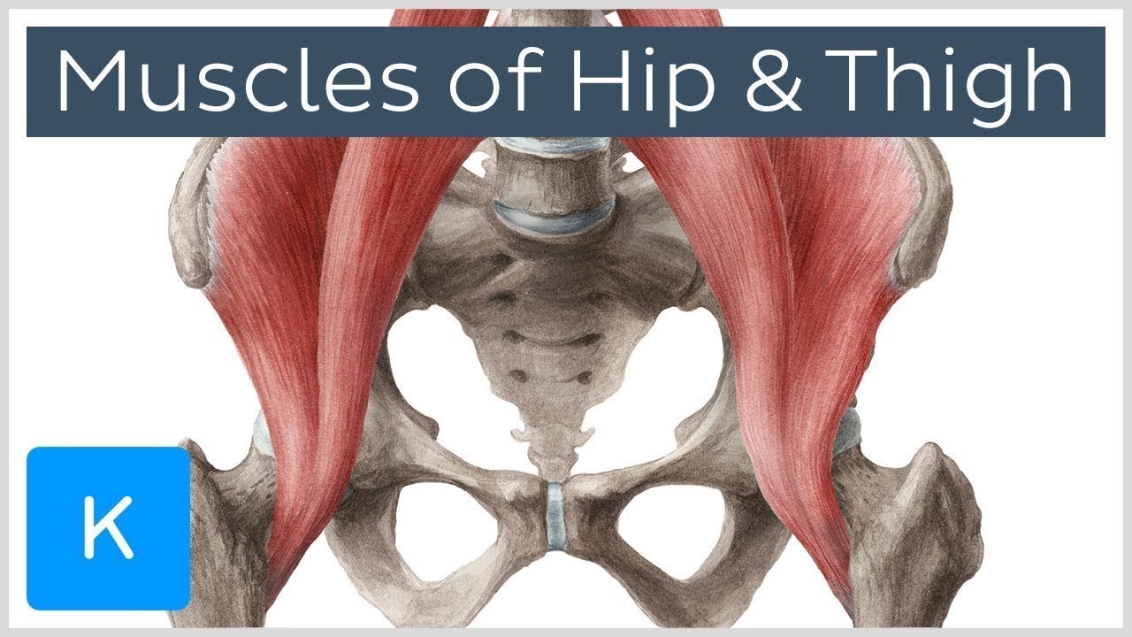
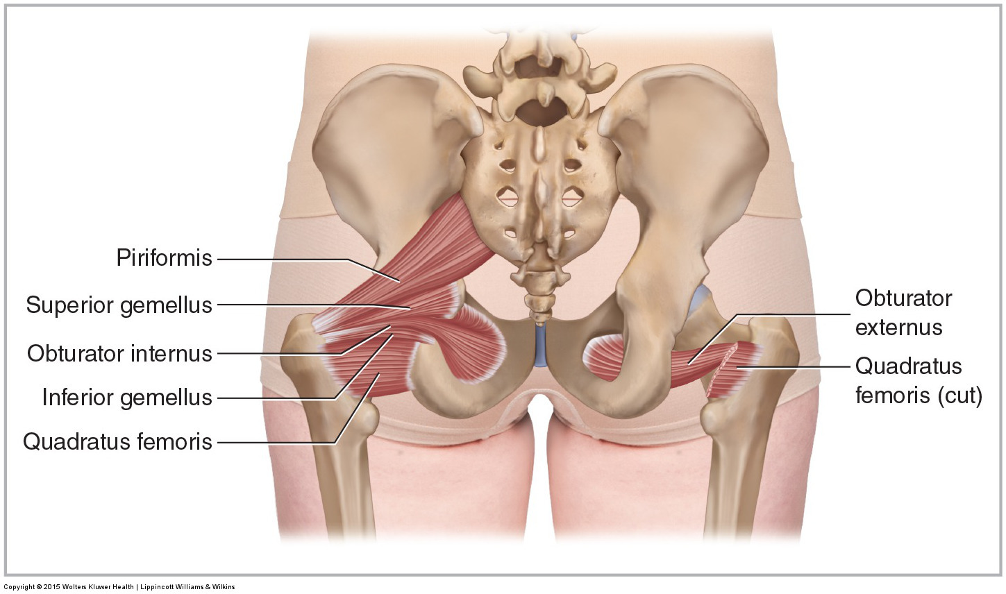
Muscles Of The Hip And Thigh Human Anatomy Kenhub Youtube from i.ytimg.com These muscles work together to flex your hip and to stabilize your hip and lower back during activities such as walking, running, and rising from a chair. Small and deep muscles which mainly externally rotate the thigh at the hip joint and stabilize the pelvis. Related online courses on physioplus. These muscles include the gluteus maximus muscle (the largest muscle in the body) and the hamstrings group, which consists of the biceps femoris, semimembranosus, and semitendinosus muscles. The hip joint allows for movement in three major axes, all of which are perpendicular to one another. It's formed by the joining of three muscles: The hip joint is a ball and socket synovial joint, formed by an articulation between the pelvic acetabulum and the head of the femur. The quadriceps muscles are four powerful muscles at the front of the thigh involved in movement.
Learning the anatomy of your hip will better enable you to pinpoint your pain and work with your doctor to keep it from limiting your life.
The different anatomical areas of the gluteal region:. See anatomy hip muscles stock video clips. The adductors all originate on the pubis and insert on the medial, posterior surface of the femur, with the exception of the gracilis which inserts just below the medial condyle of the tibia. Adductor muscles of the hip the adductor brevis, adductor longus, adductor magnus, pectineus, and gracilis make up the adductor group. The iliacus muscle, the psoas major muscle, and the psoas minor muscle. In conclusion, a thorough understanding of pelvic and hip anatomy is important for. Muscle anatomy amazon 12 photos of the muscle anatomy amazon amazon muscle anatomy poster, muscle anatomy amazon, muscle anatomy model amazon, muscle trigger point anatomy amazon, human muscles, amazon muscle anatomy poster, muscle anatomy amazon, muscle anatomy model amazon, muscle trigger point anatomy amazon The muscles you probably know the best are your glutes. Of the quadriceps muscles, it has the least affect on flexion of the knee. Advanced hip flexor muscle anatomy. A joint capsule is a watertight sac that surrounds a joint. Pick which works for you and then. Anatomy 3d atlas allows you to study human anatomy in an easy and interactive way.
In the hip, the joint capsule is formed by a group of three strong ligaments that connect the femoral head to the acetabulum. The different anatomical areas of the gluteal region:. These muscles work together to flex your hip and to stabilize your hip and lower back during activities such as walking, running, and rising from a chair. A bursa that sometimes causes problems in the hip is sandwiched between the bump on the outer hip (the greater trochanter) and the muscles and tendons that cross over the bump. The iliopsoas muscle is a major mover of your hip joint.
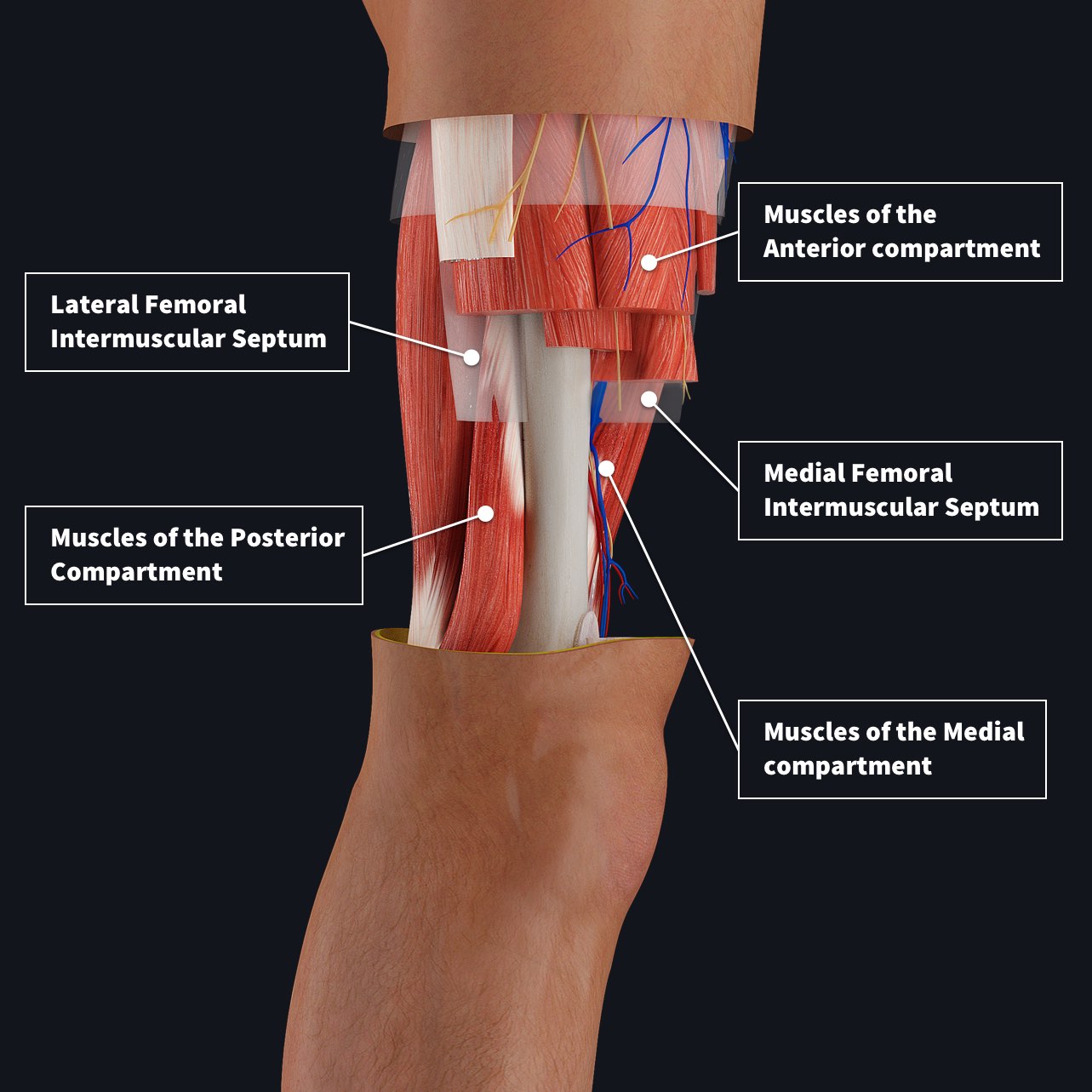
Muscle Compartments Of The Thigh Complete Anatomy from 3d4medical-cdn.s3-us-west-1.amazonaws.com Advanced hip flexor muscle anatomy. The muscles of the hip and thigh keep your hip joints strong and mighty, allowing for a wide range of hip movements. Related posts of muscles of the lower back and hip diagram muscle anatomy amazon. The iliopsoas muscle is a major mover of your hip joint. The gluteus medius muscle helps abducts the thigh along with the gluteus maximus, but can rotate the thigh inward where the gluteus maximus rotates the thigh outward. These muscles include the gluteus maximus, hamstrings muscle group consisting of the biceps femoris, semimembranosus, and semitendinosus, and the adductor magnus. Pelvis and acetabulum, with muscle attachment sites. The muscles you probably know the best are your glutes.
Anatomy 3d atlas allows you to study human anatomy in an easy and interactive way.
The hip's essential muscles are the sartorius, rectus femoris, gluteus minimus and medius, iliopsoas, adductors, and hamstrings. The anatomy of the hip and back is comprised of numerous parts that can be injured or wear out, and many problems that occur in this area can display the exact same symptoms or pathology. Thigh magnetic resonance imaging the thigh has some of the body's largest muscles. One at the left hip, and one at the right hip. Muscle anatomy amazon 12 photos of the muscle anatomy amazon amazon muscle anatomy poster, muscle anatomy amazon, muscle anatomy model amazon, muscle trigger point anatomy amazon, human muscles, amazon muscle anatomy poster, muscle anatomy amazon, muscle anatomy model amazon, muscle trigger point anatomy amazon Left hip muscles anatomy : Left hip muscles anatomy : The hip joint is a ball and socket synovial joint, formed by an articulation between the pelvic acetabulum and the head of the femur. The muscles are broken down into three layers, and are primarily used to assist with the breathing process. Related online courses on physioplus. In human anatomy, the muscles of the hip joint are those muscles that cause movement in the hip. In utero fetal hips lie typically in flexion, abduction and external rotation, with the left hip usually muscular anatomy. A joint capsule is a watertight sac that surrounds a joint.
Hip muscle strains can improve with home treatment, but severe strains may need. Now that you watched the video, you shou. The anatomy of the hip and back is comprised of numerous parts that can be injured or wear out, and many problems that occur in this area can display the exact same symptoms or pathology. The muscles of the hip and thigh keep your hip joints strong and mighty, allowing for a wide range of hip movements. The hip joint is a ball and socket synovial joint, formed by an articulation between the pelvic acetabulum and the head of the femur.
Muscles Of The Pelvis from learnmuscles.com These muscles include the gluteus maximus, hamstrings muscle group consisting of the biceps femoris, semimembranosus, and semitendinosus, and the adductor magnus. Use the mouse scroll wheel to move the images up and down alternatively use the tiny arrows (>>) on both side of the image to move the images.>>) on both side of the image to move the images. The adductors all originate on the pubis and insert on the medial, posterior surface of the femur, with the exception of the gracilis which inserts just below the medial condyle of the tibia. The hip joint connects the lower extremities with the axial skeleton. The posterior muscle group is made up of the muscles that extend (straighten) the thigh at the hip. The different anatomical areas of the gluteal region:. Thigh magnetic resonance imaging the thigh has some of the body's largest muscles. The gluteal muscles consist of the gluteus maximum, gluteus medius, and gluteus minimus.
The iliacus muscle, the psoas major muscle, and the psoas minor muscle.
Of the quadriceps muscles, it has the least affect on flexion of the knee. Discover the muscle anatomy of every muscle group in the human body. The hip joint connects the lower extremities with the axial skeleton. Related online courses on physioplus. Your email address will not be published. Anatomy of the muscular system. The posterior muscle group is made up of the muscles that extend (straighten) the thigh at the hip. The iliacus muscle, the psoas major muscle, and the psoas minor muscle. The muscles are broken down into three layers, and are primarily used to assist with the breathing process. In human anatomy, the muscles of the hip joint are those muscles that cause movement in the hip. It's formed by the joining of three muscles: In conclusion, a thorough understanding of pelvic and hip anatomy is important for. Thigh magnetic resonance imaging the thigh has some of the body's largest muscles.